Saturday, March 26, 2011

Photosynthesis: Environmental Factors

Rate of photosynthesis:
Measured by the net CO2 uptake: photosynthetic uptake – photorespiratory evolution – respiratory evolution

There are 4 main factors affecting the rate of photosynthesis in plants:

1) Light Intensity
Generally, as light intensity increases, the rate of photosynthesis also increases.
When there is no light, net CO2 production is negative due to cellular respiration.
Light-compensation point - when the amount of CO2 used is equal to the CO2 produced: net CO2=0
Light Limited phase - For sometime, the rate of photosynthesis increases in direct proportion to light (the relationship is linear). The reaction is speeding up as substrate concentration is increasing until the enzymes are working at full potential.
Light Saturation point - carbon fixation has reached maximum rate: enzyme concentration in limiting.

2) Temperature
Between 10-30 degrees: rate increases with temperature.
40 + degrees: the enzymes which catalyze the reactions within photo synthesis become denatured and are no longer able to function. This causes the rate to decrease after this point.

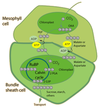
3) Carbon dioxide Concentration
Generally, the higher the concentration of CO2 in the atmosphere, the higher the rate of photosynthesis.
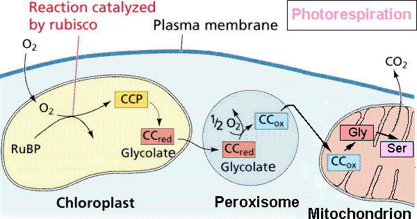
The enzyme rubisco involved in the Calvin Cycle also has a high affinity for oxygen. The greater the carbon dioxide concentration, the less chance RuBP will oxidized.
Once again, this increase in rate of photosynthesis is only until the enzymes involved are working at full potential.

4) Water Concentration
Water is another reactant in photosynthesis, and therefore causes an increase in reaction rate also until the enzymes are working at their full potential.

Can a plant get too much water?

Yes, in large plants, root cells are responsible for carrying water to upper cells for photosynthesis. They, themselves, perform cellular respiration to survive which requires oxygen which they absorb from air pockets in the soil. If the soil is completely saturated with water, no oxygen will be absorbed by these cells, and they will die. This may eventually lead to the death of the entire plant.

Photosynthesis: Alternative Mechanisms

The type of photosynthesis discussed in the last blog is referred to as C3 photosynthesis. Some plants have slightly modified mechanisms of carbon fixation due to a particularly dry climate.

Photorespiration- The oxidation of RuBP:

How it happens?
When it is warm, plants close their stoma to save water.
CO2 cannot enter the leaf.
Oxygen concentrations increase.
Oxygen, instead of Carbon dioxide, then bonds to RuBP.

The result?
Less carbohydrates are produced by photosynthesis since PGA molecules are removed— instead of 2 PGAs which are produced during photosynthesis, only one PGA and another 2 carbon glycolate are formed from RuBP.

How is it overcome in hot climates?

C4 photosynthesis
The blow is a good summary of C4 photosynthesis:

Overall, CO2 is added to a 3-carbon PEP molecule forming a 4-carbon OAA, and then malate, which then passes into the bundle sheath cells where the CO2 can be released and used for photosynthesis.

Location: Cytoplasm of mesophyll cell/bundle sheath cell. (spatial separation)
Note:
C3 Photosynthesis: 18 ATP used per glucose molecule.
C4 Photosynthesis: 30 ATP used per glucose molecule.

Crassulacean acid metabolism (CAM)
The below is a good summary of CAM:

Overall, during the night, CO2 is stored in the vacuoles of the plant in the form of organic acids. During the day, this CO2 is released and used for photosynthesis.

Location: Cytoplasm (all in one place; different from C4 photosynthesis - Temporal Separation: day/night)

Wednesday, March 23, 2011

Photosynthesis: Calvin Cycle

Although the Calvin Cycle does not rely directly on exposure to the sun for energy, its enzymes still require sunlight to be activated. This cycle follows the light reactions and is where the carbohydrates are synthesized.

The bellow diagram illustrates the process well.


It is divided into 3 phases:

1)Carbon fixation
3 carbon dioxide + 3RuBP -> unstable 6 carbon compound -> 3 PGA
Enzyme : Rubisco (Most abundant protient on earth. Also large.)

2)Carbohydrate synthesis
3PGA + 6ATP -> 6 1,3-bisphosphoglycerate + 6ADP
Enzyme: PGA kinase
1,3-bisphosphoglycerate + 6NADPH -> 6G3P + 6 Pi + 6NADP+
Enzyme : G3P dehydrogenase
*Note : 1G3P -> SUGAR

3)Ribulose biphosphate (RuBP) regeneration
5G3P + 3ATP -> 3RuBP + 3ADP + 2Pi

Overview:
Net ATP: -9
Location: Stroma
Reactants : 3 RuBP + 3CO2 + 9ATP + 6 NADPH + 5 H2O
Products: 9 ADP + 8Pi + 6 NADP+ + G3P + 3 RuBP

Tuesday, March 22, 2011

Photosynthesis: Cyclic Electron Flow

In some cases, the electrons may take an alternate cyclic pathway in which pigments capture energy from the sun in photosystem 1.


As per usual, the energy is transferred to the alpha glucose in the reaction centre and an electron is transferred to the primary electron acceptor.

From there, ferredoxin carries it to the b6-f complex and plastocyanin carries it back to photosystem 1 where it may replace another lost electron.

In the process, a proton gradient is created for ATP synthesis.

This type of electron flow DOES NOT allow for carbon fixation because there are no electrons released to form NADPH, a energy carrier necessary for the reduction of carbon dioxide.

Sunday, March 20, 2011

Photosynthesis: Chemiosmosis (Chloroplast vs. Mitochondria)


Mitochondria:

- electrons from organic substances
- chemical energy transferred to ATP
- electrons pumped from matrix to inner membrane space
- ATP goes into matrix

Chloroplast:

- electrons from water
- light energy transferred to ATP
- protons pumped from stroma to lumen (thylakoid space)
- ATP goes into stroma

Photosynthesis: Light Reactions

What is it?
Photosynthesis is the process by which autotrophs convert light energy into potential chemical energy which can then be used by both themselves and heterotrophs.

Where does it take place?
Photosynthesis occurs in the chloroplasts of plants which are usually located in the mesophyll tissues of their leaves.
To the right is an image of a chloroplast. Refer to it as we walk through the entire process.
Chlorophyll is the green pigment that absorbs the light energy.

Below is the electromagnetic spectrum. Visible light (from 380-750) is what powers photosynthesis. More specifically, the pigments responsible for capturing light absorb violet, blue and red light best. Green is reflected.

Embedded in the thylakoid membrane are numerous photosystems. These consist of light harvesting complexes containing chlorophyll-b,and a reaction-center complex containing 2 chlorophyll-a's and an electron acceptor.
When light is absorbed by electrons in the chlorophyll are excited. The energy is transferred from chlorophyll-b molecules to the chlorophyll-a molecule from which the excited electron can be ejected and captured by the electron acceptor.

In photosystem 2, the electron that is lost from the a-chlorophyll is replaced by an electron obtained from the splitting of water into oxygen and hydrogen ions. The hydrogen ions remain in the lumen and contribute to the charge buildup.

From the primary electron acceptor, the electron proceeds through a transport chain consisting of plastoquinone, a cytosome complex, and plastocyanin. In this process, more H+ are pumped into the lumen allowing for ATP synthesis using an ATP synthase structure similar to the one used in cellular respiration.
Note: 4H+ yield 1ATP

At the end, this electron is passed to photosystem 1 where it replaces an excited electron that has been used to create NADPH.

This process is summarized well in the diagram below.

Overview
Net ATP: 24
Location: Thylakoid Membrane
Reactants : 48 Photons + 12 water + 24ADP + 12NADP+
Products: 6O2 + 24ATP + 12NADPH

Saturday, March 19, 2011

Fermentation

Fermentation occurs when there is not enough oxygen present (in anaerobic conditions).
In such conditions, only glycolysis may proceed, but the pyruvate is unable to enter the other components of cellular respiration. The 2 ATPs gained by glycolysis alone is enough to sustain simple life forms, like yeast.

Glycolysis requires the presence of NAD+ which only exists in limited quantities within cells. If this supply runs out, glycolysis can no longer proceed, and the cell will no longer have a source of energy. To overcome this, cells have developed the process of fermentation.

There are 2 types of fermentation: alcoholic fermentation (done by yeast cells) and lactic acid fermentation (done by animal cells)

Alcoholic Fermentation:
In alcoholic fermentation, pyruvate is made into ethanol. Ethanol is the final electron acceptor. In the process, NAD+ is changed back to NADH allowing the cycle to continue.

Humans take advantage of alcoholic fermentation to create alcoholic beverages.


Lactic Acid Fermentation:
In lactic acid fermentation, pyruvate is made into lactate. Lactate is the final electron acceptor. In the process, NAD+ is changed back to NADH allowing the cycle to continue.

Lactic acid fermentation occurs in human muscles cells during a work out when not enough oxygen is present to meet the demands of the cells. This causes the burn.


Cellular Respiration: Oxidative Phosphorylation

The below diagram is a good summary of what happens during oxidative phosphorylation (including the electron transport chain and ATP synthesis)
During this process, electrons are passed to a series of enzymes via electron carriers.
It is first given to NADH dehydrogenase, then carried to the cytochrome b-c complex by ubiquinone, then carried to the cytochrome oxidase complex by cytochrome c. Here, oxygen acts as the final electron acceptor. These enzymes are in order of increasing electron affinity.
For every electron passed at each of the enzymes, energy obtained is used to transport a H+ ion into the inner membrane space of the mitochondria.
This creates potential energy in the form of positive charge build up.
Finally, these protons enter the matrix through the ATP synthase structure creating one ATP molecule for every H+ that was pumped out.


Why is the net ATP production of cellular respiration 36-38?
This is because, as can be seen from the overall diagram of cellular respiration on the right, the NADHs that are produced during glycolysis must be brought into the matrix of the mitochondria. Passage through the outer membrane is simple, however, they cannot pass through the inner membrane without some help.
There are 2 ways this occurs: through aspartate or glycerate phosphate.
When aspartate is used, the NADH passes through as NADH.
When glycerate phosphate is used, the electron passes through and attaches to FAD to make FADH2.

RECALL:
NADH enters at first enzyme of ETC pumping 3 electrons, creating 3 ATPS.
NADH enters at second enzyme of ETC pumping 2 electrons, creating 2 ATPS.

This is why there may be slight variations.

Reactants: 6NADH (from Krebs), 2NADH (from pyruvate oxidation), 2FADH2 (from Krebs), 2FADH2 OR 2NADH (from Glycolysis), 32-34 ADP, 32-34 phosphate groups, 6Oxygen molecules, 12H+
Products: 8-10NAD+, 2-4FAD+, 24H+, 32-34 ATP, 6Water molecules
Location: Inner membrane of the mitochondria
Net ATP: 32-34

Cellular Respiration: Krebs Cycle

The bellow diagram serves as a good summary for what occurs during the Krebs Cycle.


By the end of the Krebs cycle, all the carbons from the initial glucose molecule have been expelled as carbon dioxide. (2 during pyruvate oxidation, and 4 during the krebs cycle)

Energy that has been stored in the electron carrier molecules (NADH and FADH2) can now proceed to the electron transport chain where they can be used to synthesize ATP; the for of usable energy in cells.

Reactants: 2Acetyl-CoA, 6NAD+, 2ADP, 2 phosphate groups, 2FAD
Products: 2CoA, 4carbon dioxide, 6NADH, 6H+, 2FADH2, 2ATP
Location: Mitochondrial matrix
Net ATP: 2

Cellular Respiration: Pyruvate oxidation

During pyruvate oxidation, the two pyruvates are transported into the matrix of the mitochondria. A transport protein is required to allow for its passage through the inner membrane.

First, a low energy carboxyl group is removed as carbon dioxide.
*This is a decarboxylation reaction catalyzed by the enzyme: pyruvate decarboxylase.
Secondly, NAD+ oxidizes the 2 carbon compound forming NADH.
Thirdly, sulfur containing CoA attaches to the acetate component.

The acetyl-CoA is now ready for the Krebs Cycle!

Reactants: 2 pyruvate, 2NAD+, 2CoA
Products: 2 acetyl-CoA, 2NADH, 2H+, 2Carbon dioxide molecules
Location: Mitochondrial Matrix
Net ATP: none

This video also illustrates the electron transport chain discussed in the next blog as well!

Cellular Respiration: Glycolysis

The first phase of cellular respiration is called glycolysis. Only 2.2% of the energy from cellular respiration is attained through glycolysis.
The above image illustrates the process of glycolysis in detail. It includes the names of the enzymes responsible for catalyzing each individual reaction. It also included the names and structural diagrams of the intermediate reactions.

In general, gylcolysis can be divided into 2 phases: energy investment (Step#1-3) and energy payoff (Step#4-10). During the energy investment phase, 2 ATP molecules are used. In the energy payoff phase, 4 ATPs in addition to 2 NADHs are synthesized.

Reactants: glucose, 2ATP, 2NAD+, 4ADP, 2 phosphate groups
Products: 2 pyruvate, 4ATP, 2ADP, 2H+, 2NADH
Location: Cytosol
Net ATP: 2

This video explores glycolysis with particular focus on the enzymes involved.

Enzymes

Enzymes are biological catalysts that speed up chemical reactions within the body without being consumed. They can be used over and over again.
All reactions require an initial input of energy, called activation energy. Enzymes lower this activation energy as shown in the diagram to the left.

Key terms:
Substrate - the reactants which the enzyme acts on.
Enzyme-substrate complex - the complex formed when the substrate molecules bind to the enzyme.
Active site - where the substrates bond to the enzymes
Cofactors - non-organic enzyme helpers
Coenzymes - organic enzyme helpers
Allosteric site - another region on the enzyme where materials can bond to the enzyme locking it in either its active or inactive shape.
Feedback inhibition - when the product of the reaction bonds to the allosteric site of an enzyme locking it into its inactive form.

The cycle:
The below image is a good summary of how the catalytic cycle works.

Inhibition:
There are two types of inhibition: competitive and non competitive.
Competitive - inhibitor competes with substrate to bind to the active site of the enzyme.
Non-competitive - inhibitor binds to the allosteric region of enzyme.

Here is a video showing the importance of enzymes in out body.

Sunday, March 6, 2011

Methods of Transport: Active

Active transport in cells requires the use of energy to transport products against the concentration gradient. 30-40% of all energy within a cell is used for transport.

Individual molecules:
Transport proteins actively pump molecules in/out of the cell using energy.
An example of such a pump would include the sodium-potassium pump. Three sodium ions within the cytosol bind to the protein causing it to change shape. The protein can then attain a phosphate group from ATP causing another change in shape that releases the sodium ions outside the cell, and allowed potassium ions to bond to the protein. The phosphate group then dislocates making the protein return to its original shape releasing the potassium ions within the cell.

Cells like to have a high concentration of potassium ions within them, and a low sodium ion concentration. To allow this to work,


In Bulk:
In bulk, material is moved into/out of a cell using endocytosis and exocytosis.


Endocytosis:
Involves the movement of material into the cell through the creation of new vesicles from the cell membrane.
When liquids are brought in, it is called, pinocytosis.
When solids are brought in, it is called, phagocytosis.

Exocytosis:
Involves the movement of material out of the cell through fusing vesicles with the plasma membrane.

Methods of Transport: Passive

Passive transport does not require the use of energy to move material into/out of the cell.
There are two types of passive transport: osmosis and diffusion

Osmosis:
Osmosis is a special type of diffusion; Movement of water along a concentration gradient until equilibrium is established.
In isotonic solutions, the concentration of solute is equal in both the inside and out side of the cell. Therefore, there is no net change in movement of water. This environment is ideal for animal cells. When plant cells are placed in isotonic solutions, however, they are called 'flaccid' and are not as supported by the cell wall.
In hypertonic solutions, the concentration of solute outside the cell is greater. Therefore, solvent moves out of the cell causing it to shrivel. Animal cells become shriveled and plant cells become plasmolyzed.
In hypotonic solutions, the concentration of solute less than inside the cell. Therefore, solvent moves into the cell causing it to swell. This is ideal for plant cells as they become turgid. But animal cells may become lysed and burst.

Diffusion:
Diffusion is defined as the movement of solute molecules from an area of high concentration to an area of low concentration (along a concentration gradient) until equilibrium is established.

Diffusion in cells can be facilitated. This involves the use of transport proteins to move these solute molecules across the membrane. It is still considered passive transport because the particles are moving without the use of energy according to the concentration gradient.